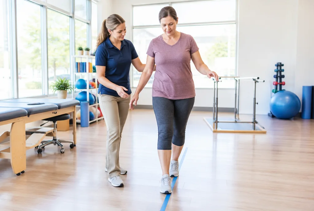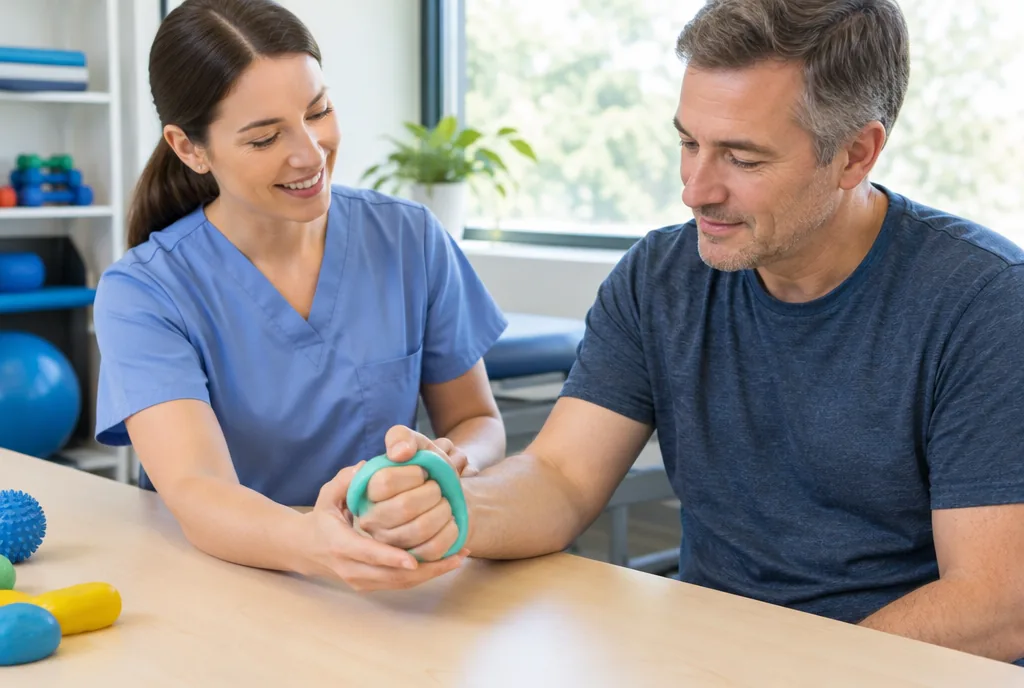
Foot drop exercises can strengthen ankle dorsiflexion, preserve range of motion, and make walking practice more purposeful, but they do not reveal why the front of the foot will not lift. That distinction matters: a sudden foot drop needs medical evaluation before a home program begins, because the cause can change the rehabilitation plan. Once the cause is understood, specific work on toe lifting, calf flexibility, balance, and walking can give each practice session a clear job.
Understand what foot drop can signal before you exercise
Foot drop is the inability to lift the forefoot, or dorsiflex it, because the muscles that lift the foot are weak or paralyzed. Those muscles include the tibialis anterior, extensor hallucis longus, and extensor digitorum longus. The visible problem may be a dragging toe, but the source can be in different places along the nervous system.
Central causes include lesions of the motor cortex, such as stroke and cerebral palsy. Injury within the spine can involve the L4, L5, S1, or S2 nerve roots. A peripheral mononeuropathy of the peroneal nerve can also cause foot drop. The common peroneal nerve branches from the sciatic nerve, passes around the outside of the fibular head, and divides there; that exposed route makes it particularly vulnerable to pressure.
Causes named by neurologic and clinical sources include multiple sclerosis, spinal muscular atrophy, ALS, Charcot-Marie-Tooth disease, diabetic neuropathy, herniated disk, spinal stenosis, knee injury, prolonged hospitalization, and compression from a cast. Even prolonged cross-legged sitting can compress the peroneal nerve at the fibular head. A new or sudden change should be assessed by a doctor rather than treated as a routine exercise problem.
Foot drop exercises begin with ankle dorsiflexion
Start where the movement is easiest to see: seated with the knee bent and the heel grounded. Slowly pull the forefoot and toes toward the shin, then return with control. This active ankle dorsiflexion can be practiced for 2 to 3 sets of 20 to 30 repetitions on each side. A small movement still gives useful practice; repetition is the point.
Use elevated toe taps to give the toes room to move
Place the heel on a step so the toes hang over the edge. Let the forefoot lower slowly, then actively lift the toes toward the ceiling. Perform up to 20 to 30 repetitions for 2 to 3 sets. The heel stays supported while the ankle moves through the available range.
Let the stronger side assist toe raises
For assisted toe raises, place the unaffected foot beneath the toes of the affected foot and help them upward. Begin with 2 sets of 10 repetitions. A useful variation is the negative: use the hand or stronger side to bring the foot into maximum dorsiflexion, then lower it slowly into plantarflexion over 3 to 5 seconds. When assisted raises become easier, the progression is active dorsiflexion without help and then light resistance.
Trace the alphabet when the ankle needs more movement variety
While seated or lying down, use the big toe to draw the alphabet in the air. The ankle alphabet works the ankle through a broad range of motion without turning the session into a long workout. It pairs naturally with the more focused toe-lifting drills above.
Build control with a resistance band and calf stretch
Add a resistance band only after the foot can move with control. Sit with the knee at 90 degrees, loop the band around the forefoot, and secure it under the foot or at a fixed point. Pull the foot back toward the shin against the band. For eversion, turn the outside of the foot outward against resistance; this targets the peroneal muscles.
Strength work needs flexibility alongside it. In a gastrocnemius calf stretch, stand in a lunge with the back leg straight and the heel on the floor. Hold 30 to 60 seconds, repeat 3 to 5 times on each side, and practice twice daily. For the soleus, use the same setup with the rear knee bent. Regular calf stretching matters because absent dorsiflexion can contribute to Achilles tendon shortening and an equinus contracture.
Earn gait practice with balance training first
Foot clearance is only part of safe walking. Before moving on to gait drills, you should be able to stand safely on one leg for 3 sets of 20 seconds on each side. This checkpoint is often missed in home programs, even though balance control is needed every time the body passes over one leg during a step.
Use a tandem stance by placing one foot directly in front of the other and holding for 30 seconds. Progress in a deliberate order: hold a chair first, then use only a light touch, then stand freely, and finally try closed eyes. More challenging reactive balance work, such as gentle perturbations from a therapist, belongs in supervised therapy rather than an unsupervised home routine.
Walk with foot drop without ignoring the fall risk
It can be appropriate to practice walking, but the gait pattern and support matter. Without correction, foot drop may produce a high-stepping gait: the hip and knee lift excessively so the toes clear the ground. It may also produce a foot slap when the foot lands. Both patterns can make walking more demanding, and toes can catch on rugs, curbs, or uneven surfaces.
One simple cue is “heel first”: consciously place the heel down first, then control the rest of the step. This cue does not diagnose the cause or replace an individualized gait plan. It gives practice a specific focus while a clinician determines which exercises, brace, or other support fit the person’s movement.
Choose an AFO for foot drop by the movement you need
An AFO for foot drop—an ankle-foot orthosis—holds the foot in a neutral position to help prevent catching. There is no single best AFO. Selection depends on the cause and severity of foot drop, ankle stiffness, spasticity, knee control, activity level, and shoe choice.
| AFO type | How it is described | When it may fit |
|---|---|---|
| Solid AFO | Rigid, with no ankle joint movement. | Severe paralysis, spasticity, or instability. |
| Articulated AFO | A hinge allows controlled plantarflexion and dorsiflexion with adjustable stops. | When some movement is useful but spasticity also needs control. |
| Posterior leaf spring AFO | A light, flexible carbon or plastic spring. | Mild, isolated foot drop without side-to-side instability; it can fit regular shoes. |
| Ground-reaction AFO | Influences knee stability as well as the ankle. | Quadriceps weakness or crouch gait; it is bulkier. |
Consider functional electrical stimulation when nerves are intact
Functional electrical stimulation (FES) delivers electrical impulses to the peroneal nerve or directly to the tibialis anterior to trigger dorsiflexion during the swing phase of walking. It can serve a therapeutic role, by training movement, or an orthotic role, by supporting foot lift during gait.
FES is especially suited to people whose peripheral nerves remain intact, including some people after stroke or with multiple sclerosis. It is not effective after a complete peripheral nerve transection. For people who are appropriate candidates, research in people with stroke and foot drop has reported improvements in walking speed, endurance, and coordination after FES use.
Separate common exercise myths from realistic recovery
Myth: More exercise can identify the cause. Exercises can work on mobility and control, but they cannot distinguish a nerve compression from a stroke, spinal involvement, or another cause. Sudden foot drop should be medically evaluated before starting a home program.
Myth: Every foot drop recovers on the same schedule. Prognosis follows the cause and severity. Neurapraxia, a mild nerve injury, usually recovers completely in about 3 months. With axonotmesis, where axons are damaged but the nerve sheath remains intact, recovery may take much longer and may not be complete. Nerves regrow at roughly 1 to 3 millimeters per day, so recovery can take months, and over longer distances even years.
Myth: A brace means exercise has failed. An AFO and an exercise plan solve different parts of the problem. A well-matched brace can hold the foot neutral for safer walking, while exercises address dorsiflexion, flexibility, and balance. In traumatic or nerve-related cases, recovery may be partial or complete; in progressive neurologic diseases, foot drop usually remains permanent.
Frequently Asked Questions
Can foot drop be corrected with exercise?
Exercise can improve mobility, strength, and balance, but it cannot establish the cause of foot drop. Whether foot drop improves depends on the underlying condition and severity of nerve injury.
What exercises help foot drop?
Useful options include active ankle dorsiflexion, elevated or assisted toe raises, slow negative toe raises, resistance-band dorsiflexion and eversion, calf stretches, the ankle alphabet, and balance training.
Is it good to walk with foot drop?
Walking practice should account for toe catching and fall risk. A heel-first cue, appropriate balance preparation, and an AFO when indicated can help make gait practice safer.
Can you recover from foot drop?
Recovery is possible, especially after some traumatic or nerve injuries. Neurapraxia commonly recovers in about 3 months, while axonal injury can take much longer and may not fully recover.
What causes foot drop?
Causes can be central, spinal, or peripheral. They include stroke, spinal nerve-root injury, and peroneal nerve injury or compression, as well as conditions such as multiple sclerosis and diabetic neuropathy.