Diffuse axonal injury (DAI) is a traumatic brain injury in which rapid acceleration, deceleration, or rotation stretches the brain’s long nerve fibers, called axons. It can cause a person to lose consciousness even when an early CT scan looks nearly normal. That mismatch is one reason DAI is so challenging: the injury can be widespread at a microscopic level, while the first images may show little. If you or someone close to you has a significant head injury, get immediate medical assessment, and discuss recovery with the treating clinical team rather than predicting it from one scan or one grade.
What diffuse axonal injury does inside the brain
Axons are the long projections that allow nerve cells to communicate across the brain. In diffuse axonal brain injury, rapid linear or rotational forces stretch and can tear axons in white matter. The name “diffuse” describes the broad distribution of injury, although the damage is multifocal rather than evenly spread through every part of the brain. Some clinicians also use the term traumatic axonal injury for that reason.
The first mechanical event is only part of the story. A complete mechanical break of an axon, called primary axotomy, is relatively uncommon. More often, partial damage interrupts axoplasmic flow, the transport system within the axon. Proteins then accumulate, and swollen segments called axonal varicosities and axonal bulbs can form. Beta-amyloid precursor protein can accumulate inside axons within two to three hours and is a sensitive histologic marker of this process.
Damage may also progress after the initial trauma. Mechanical membrane pores and voltage-gated sodium channels allow calcium to enter cells. That calcium activates calpains, enzymes that break down spectrin and other proteins in the axonal skeleton; this process can begin about 15 to 30 minutes after injury. A secondary inflammatory and apoptotic process can continue for hours to days, eventually severing additional axons. Mitochondrial injury, loss of ATP, and release of pro-apoptotic factors are part of this secondary cascade.
DAI commonly affects places especially vulnerable to shearing forces: the gray-white matter junction, particularly in frontotemporal areas; the corpus callosum, which connects the two cerebral hemispheres; and the brainstem. In one imaging description, the gray-white junction was the most frequent location at 67%, followed by the corpus callosum at 20%; the splenium and posterior body of the callosum were common callosal sites. Brainstem lesions were described in the dorsolateral midbrain and upper pons. Injury in these regions can disrupt the networks that support wakefulness and coordinated brain function.
Timing also explains why DAI can be difficult to document with ordinary tissue methods immediately after trauma. Routine H&E staining may not show the injury for about 24 hours, while silver staining can show it after 12 to 18 hours. Beta-APP immunostaining can reveal axonal injury after two to three hours. In the first week, frank demyelination is not visible; maturing oligodendrocytes decline during the first three days and recover by day seven. These laboratory details are not bedside tests, but they show that axonal injury changes over time. The fact that a process evolves after the trauma does not mean you should wait to seek care. A person with a suspected head injury needs prompt medical evaluation.
Diffuse axonal injury symptoms: why consciousness is the key clue
The chief symptom of diffuse axonal injury is loss of consciousness, often a coma lasting six hours or longer. In intensive care settings, 83.6% of patients with DAI had an admission Glasgow Coma Scale (GCS) score of 3 to 8, the range used for severe traumatic brain injury. A GCS below 8 for more than six consecutive hours supports a clinical diagnosis of DAI.
Symptoms vary with injury severity. Mild DAI may involve a brief loss of consciousness or confusion along with headache, dizziness, nausea, and fatigue. With severe DAI, a person may have no lucid interval after the trauma, abnormal pupil responses, or abnormal flexing or extending postures. These findings require urgent medical attention; they are not signs to monitor at home.
Why one symptom list cannot predict one person’s course
DAI affects networks rather than a single small brain area, so the visible effects can differ widely. The initial GCS is clinically important, but it is not the entire prognosis. Age, pupil reactivity, coma duration, blood hemoglobin level, CT classification, autonomic dysregulation, hypotension, hypoxia, and hyperglycemia have all been identified as independent prognostic factors. Repeated examination and the full medical picture are more informative than trying to draw a conclusion from one symptom.
Seek emergency evaluation after a head injury, especially if you or the injured person loses consciousness, is confused, has worsening symptoms, or shows an abnormal response. This article explains a condition; it cannot diagnose DAI or determine whether a person has it.
Why a CT scan can miss a DAI brain injury
A CT scan is important in acute head trauma, but it has real limits for DAI. Between 50% and 80% of people with DAI have an unremarkable CT scan on arrival. The scan can show small petechial hemorrhages at the gray-white matter junction, in the corpus callosum, or in the brainstem, yet fewer than 20% of patients show petechial hemorrhages on CT alone. This helps explain the unsettling but well-recognized situation in which a person is comatose while an early CT appears almost normal.
When CT does show DAI-related changes, lesions are typically 1 to 15 millimeters. In one account, 80% of patients had multiple foci on CT. But a normal or limited CT result does not rule out axonal injury. CT detects shearing injuries at a reported rate of only 20% to 50%, while MRI is about four times more sensitive.
MRI reveals much smaller signs of injury
MRI with susceptibility-weighted imaging (SWI) is the imaging method of choice for detecting DAI because it can reveal very small microhemorrhages. SWI detected microbleeds in 40% of patients in one comparison, versus 23% with gradient-echo imaging. On FLAIR sequences, lesions may appear as bright areas in white matter, particularly in the splenium of the corpus callosum.
Imaging answers an important part of the question, but it does not replace clinical assessment. Neither a single CT scan nor an MRI stage should be used alone to tell you exactly what recovery will look like. The clinical examination, GCS, CT classification, medical complications, and changes over time all belong in that conversation.
Diffuse axonal injury grades show where lesions are found
The Adams grading system began with histopathologic studies. It describes the pattern and location of axonal injury, not a fixed forecast for an individual. Gentry later translated the same general pattern into MRI-based staging: Stage I involves lobar white matter, Stage II the corpus callosum, and Stage III the brainstem.
| Grade | Defining finding | What it adds |
|---|---|---|
| Grade 1 | Microscopic axonal damage in cerebral white matter and the corpus callosum; the brainstem may occasionally be involved. | The basic axonal injury pattern. |
| Grade 2 | Grade 1 findings plus a focal lesion in the corpus callosum. | A visible callosal lesion. |
| Grade 3 | Grade 2 findings plus focal lesions in the dorsolateral quadrant of the rostral brainstem. | Brainstem involvement in addition to the other findings. |
These categories have a serious origin. The Adams classification was developed from 434 fatal traumatic brain injuries; 122 had DAI, including 10 Grade 1, 29 Grade 2, and 83 Grade 3 cases. Modern imaging can identify much smaller lesions than older pathological descriptions did. In particular, 3-Tesla MRI with SWI can detect tiny brainstem microbleeds that should not automatically be equated with the larger lesions in the original studies.
What grade 3 diffuse axonal injury can and cannot tell you
A grade 3 diffuse axonal injury includes brainstem lesions, so it is understandably alarming. In a study of 25 patients, the mean time to return of consciousness was 3.7 days for Grade 1, 12.5 days for Grade 2, and 59.5 days for Grade 3. A meta-analysis of five studies involving 258 patients found an unfavorable functional outcome in 17% of Grade 1 cases, 40% of Grade 2 cases, and 63% of Grade 3 cases.
Those are group results, not a verdict. The prognostic value of MRI grading remains disputed; smaller studies have found weak or no relationship between MRI grade and outcome. The Marshall CT classification and admission GCS are stronger predictors than DAI grade alone. Reported cases also include people with Grade 3 injury who returned to work within four months, and a 19-year-old with Grade 3 DAI and brainstem hemorrhages who regained consciousness within six weeks and reached functional independence in daily life after one year. Such cases show possibility, not a promise.
Survival and recovery depend on more than a label
There is no single survival rate that applies to every person with DAI. Across studies, mortality has ranged from 25.6% to 62%; one ICU case series reported 42%. Differences in patient populations and injury circumstances matter, and the factors present in one person’s case matter even more. For that reason, a percentage from a study should never be treated as a personal prognosis.
There are also reasons not to reduce recovery to the acute phase. In a study of 133 patients, 48.1% had a satisfactory outcome at three months, with a mean admission GCS of 9. Among survivors with pure DAI and no intracranial hematoma, almost all had a favorable outcome at 12 months, with marked improvement in the first three months. A longitudinal study found that neither clinical nor anatomical DAI findings on MRI were associated with three-year survival, Glasgow Outcome Scale results, or quality of life.
Can a person recover from diffuse axonal injury?
Yes, recovery is possible, including favorable functional outcomes years after trauma. Complete recovery can occur in very mild forms of DAI classified as concussion. Recovery after severe injury is often lengthy and variable. In one study, people with Grade 1 and Grade 2 DAI regained consciousness on average within two weeks, while Grade 3 patients took about two months. An average is not a deadline, and it does not tell clinicians what will happen in a particular case.
A useful way to frame prognosis is in stages rather than absolutes: survival through the acute injury, return of consciousness when applicable, regaining specific functions, and building independence with rehabilitation. Each stage is influenced by the person’s medical condition and response to treatment. You deserve plain explanations of uncertainty alongside realistic reasons to continue rehabilitation.
Rehabilitation turns recovery into practical goals
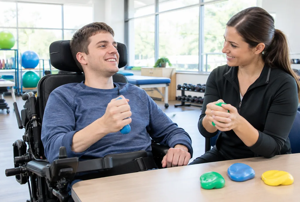
There is no operation that repairs DAI itself. Acute treatment is supportive, with attention to intracranial pressure control and prevention of secondary brain injury. Rehabilitation then brings together physical therapy, occupational therapy, speech therapy, neuropsychology, and social work. The work can be long-term after severe DAI, and it should be tailored to the person’s current abilities rather than a scan label.
Therapy can begin early, including early mobilization in the ICU when medically appropriate. A documented Grade 2 DAI case used passive and active range-of-motion exercises, balance training, functional electrical stimulation, and cognitive training, with significant improvement in the Functional Independence Measure. That is an example of a program, not a universal prescription.
- Protect and stabilize. Early care focuses on controlling intracranial pressure and avoiding additional brain injury.
- Restore movement and daily function. Physical and occupational therapy can address movement, balance, and the activities needed for everyday life.
- Address communication and thinking. Speech therapy and neuropsychology support communication and cognitive needs.
- Plan beyond discharge. Social work and the rehabilitation team help you and your family prepare for the practical demands of continued care.
Recovery is not limited to the first hospital stay. Multi-domain cognitive programs that train attention, memory, visuospatial skills, and executive function have shown improvement even when they begin nine months after injury. Rehabilitation-related neuroplasticity and functional recovery are linked to the cardiovascular intensity of activity. In one Grade 3 report, a patient entered inpatient rehabilitation on day 17, received physical, occupational, and speech therapy for two to three weeks, and then went home. The timing and setting vary; the core point is that rehabilitation is an active part of care, not an afterthought.
Four myths that can make DAI harder to understand
Myth: A normal CT means there is no serious injury
False. An early CT may be normal in 50% to 80% of DAI cases. MRI, especially SWI, is more sensitive for the small lesions associated with axonal injury. A clinician interprets imaging alongside the person’s consciousness level and neurological examination.
Myth: Grade 3 determines a person’s future
False. Grade 3 signals brainstem involvement and carries serious group-level risks, but MRI grading is not a complete prediction tool. GCS, Marshall CT classification, pupil responses, medical complications, and changes over time all add essential context.
Myth: DAI is simply a matter of axons tearing at impact
Incomplete. Mechanical forces disrupt axons immediately, but secondary biochemical injury can continue for hours to days. The modern understanding gives substantial weight to this secondary cascade rather than viewing DAI as only a momentary mechanical event.
Myth: Recovery ends when the hospital stay ends
False. The average DAI hospital stay was nine days in one study, and 58.6% of patients stayed 11 days or fewer. That brief acute-care period is not the entire recovery. Meaningful cognitive improvement has been reported even when structured programs begin months later.
Frequently Asked Questions
Can a person recover from diffuse axonal injury?
Yes. Very mild, concussion-level DAI can resolve completely, and people with more severe injuries can achieve favorable functional outcomes. The pace and extent of recovery vary, so ask the treating team to discuss prognosis using the full clinical picture.
What happens when you have diffuse axonal injury?
Rapid acceleration, deceleration, or rotation stretches axons in white matter. Interrupted internal transport can cause swelling, while secondary inflammatory and apoptotic processes may continue damaging axons for hours to days.
What is the survival rate for patients with diffuse axonal injury?
Published mortality ranges from 25.6% to 62%, and one ICU case series reported 42%. Those figures describe different study groups, not an individual’s chance of survival; age, pupils, coma duration, hypoxia, hypotension, and other factors matter.
What is the chief symptom of diffuse axonal injury?
The chief symptom is loss of consciousness, typically a coma lasting six hours or longer. Milder injury can instead involve a brief loss of consciousness or confusion with headache, dizziness, nausea, and fatigue.
Can a CT scan rule out a DAI brain injury?
No. CT can be normal on arrival in 50% to 80% of DAI cases and detects shearing injury less often than MRI. MRI with SWI is more sensitive for tiny microhemorrhages, but imaging must be interpreted with the clinical assessment.