The human brain weighs about three pounds, yet it controls movement, sensation, language, memory, emotion and the functions that keep us alive, such as breathing and heart rate. Knowing how it is organized helps explain why a stroke or brain injury in one place causes weakness of an arm, while one a few inches away affects speech, vision or memory, and why rehabilitation can restore some of what was lost. This article covers the brain’s structure, its blood supply, its development and its capacity to change.
The Brain in Numbers
| Measure | Value |
|---|---|
| Adult weight | About 1.3 to 1.4 kg (about 3 pounds), roughly 2 percent of body weight |
| Energy use at rest | About 20 percent of the body’s oxygen and energy, roughly 20 watts |
| Blood supply | About 15 percent of the blood pumped by the heart |
| Composition | About 75 to 80 percent water; about 60 percent of the dry weight is fat, much of it in myelin |
| Neurons | About 86 billion: about 16 billion in the cerebral cortex, about 69 billion in the cerebellum |
| Glial cells | Roughly as many as neurons |
| Corpus callosum | About 200 million nerve fibers linking the two hemispheres |
| Cerebral cortex | 2 to 4 mm thick, deeply folded |
| Fastest nerve signals | Up to about 120 meters per second along myelinated fibers |
Neurons, Glia and White Matter
Neurons are the brain’s signaling cells. For decades textbooks said the brain had 100 billion of them; a 2009 count by Brazilian researchers, who dissolved brain tissue and counted cell nuclei, arrived at about 86 billion. Surprisingly, most of them sit in the small cerebellum at the back of the head, not in the large cerebral cortex. Each neuron connects with thousands of others through synapses, the junctions where chemical messengers pass signals on.
Glial cells, roughly as numerous as neurons, support and protect them. One type wraps nerve fibers in myelin, a fatty insulating layer that speeds up signals. Gray matter, on the surface of the brain and in deep clusters, contains the cell bodies of neurons; white matter, underneath, consists of myelinated fibers connecting distant regions. Damage to white matter, common in traumatic brain injury, disconnects areas that may themselves be intact, which slows thinking and processing speed.
The Two Hemispheres
The cerebrum, the largest part of the brain, is divided into left and right hemispheres, linked by the corpus callosum. Each hemisphere mainly controls the opposite side of the body. In most people the left hemisphere is dominant for language, and the right plays a larger role in spatial attention and awareness of the surroundings.
This explains typical patterns after a stroke. A left-hemisphere stroke often causes weakness of the right side and aphasia, a loss of language. A right-hemisphere stroke often causes weakness of the left side and neglect, in which the person ignores the left side of space, for example eating only from the right half of the plate.
The Four Lobes
| Lobe | Main functions | Possible effects of damage |
|---|---|---|
| Frontal | Planning, decisions, attention, personality, voluntary movement (motor cortex), speech production (Broca’s area) | Weakness on the opposite side, difficulty planning and organizing, impulsive behavior, halting, effortful speech |
| Parietal | Touch, body position, spatial awareness | Numbness, poor sense of limb position, neglect, difficulty with directions |
| Temporal | Hearing, memory, language comprehension (Wernicke’s area) | Memory problems, fluent but empty speech with poor comprehension |
| Occipital | Vision | Loss of part of the visual field, difficulty recognizing objects |
The frontal lobes, just behind the forehead, are especially vulnerable in car crashes and falls, which is why changes in behavior and judgment are common after traumatic brain injury.
Deep Structures
- Thalamus: relays sensory information to the cortex; damage can cause numbness or pain on one side.
- Hypothalamus: regulates hormones, body temperature, hunger and thirst.
- Hippocampus: forms new memories; it is sensitive to lack of oxygen, so memory problems are typical after cardiac arrest.
- Amygdala: processes fear and emotion.
- Basal ganglia: help control movement; they are affected in Parkinson’s disease.
Cerebellum and Brainstem
The cerebellum, tucked beneath the back of the cerebrum, coordinates movement, balance and fine motor skills. Damage causes ataxia: unsteady walking, tremor when reaching and slurred speech. The brainstem, made up of the midbrain, pons and medulla, connects the brain to the spinal cord and controls breathing, heart rate, blood pressure, swallowing and wakefulness. Injuries here can be life-threatening and can affect consciousness.
Blood Supply and Protection
Arteries and the Circle of Willis
Four arteries supply the brain: two internal carotid arteries in the front of the neck and two vertebral arteries along the spine. At the base of the brain they join in a ring called the circle of Willis, which can partly reroute blood if one artery narrows. A stroke occurs when one of the branches is blocked or bleeds.
Why Oxygen Matters
The brain stores almost no energy. When blood flow stops, consciousness is lost within about 10 seconds, and brain cells begin to be permanently damaged after about four to six minutes without oxygen. That is the mechanism of anoxic brain injury after cardiac arrest, drowning or choking, and the reason bystander CPR matters.
Barriers and Cushions
The blood-brain barrier, formed by tightly sealed capillary walls, keeps many substances in the blood out of brain tissue, which also makes some medications hard to deliver. About 150 ml of cerebrospinal fluid, renewed several times a day, cushions the brain inside the skull. Three membranes, the meninges, and the skull itself provide further protection.
How the Brain Develops
Most neurons are produced before birth. After birth the brain grows fast: it roughly doubles in size in the first year and reaches about 90 percent of adult volume by age six, mainly through new connections and myelin. The prefrontal cortex, which handles judgment and impulse control, keeps maturing into the mid-20s, one reason brain injury in teenagers and young adults can affect development of these skills.
Neuroplasticity: How the Brain Recovers
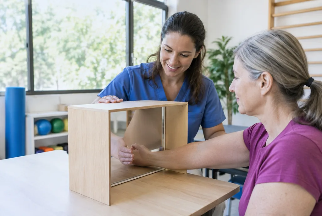
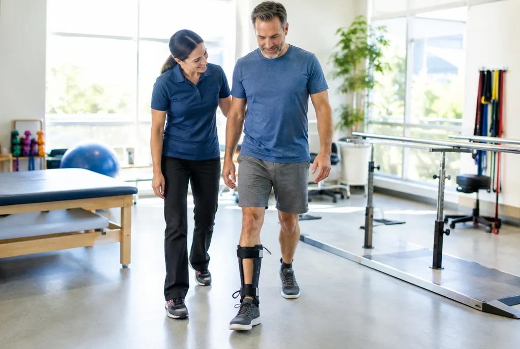
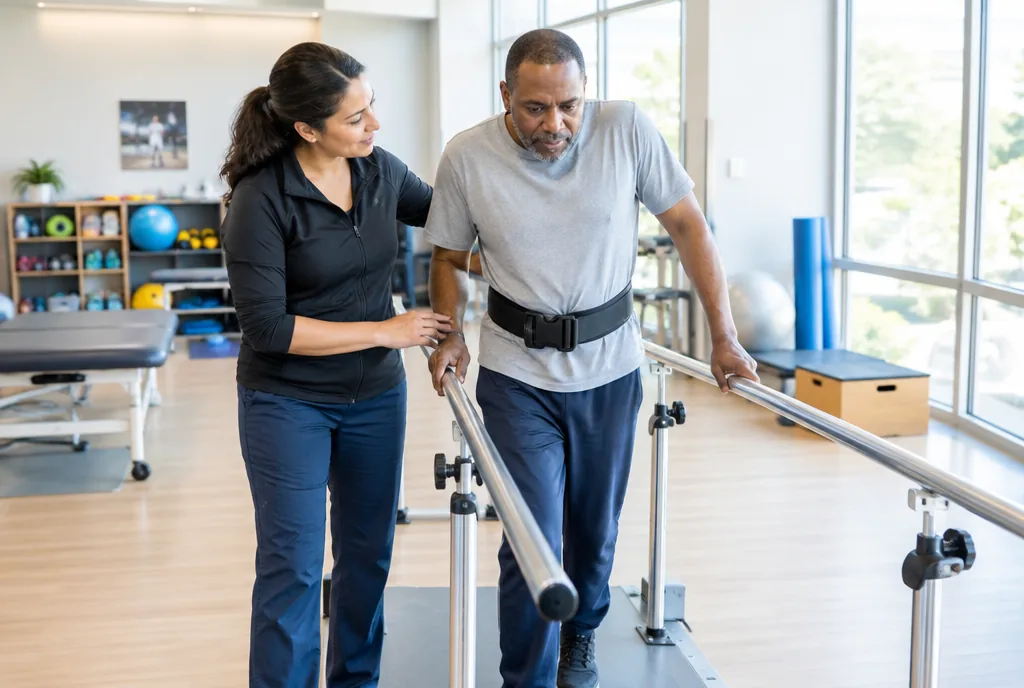
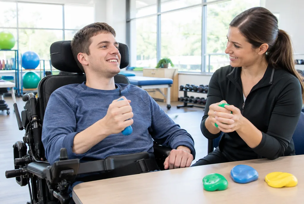
The brain changes its connections throughout life in response to experience and practice. After an injury, dead neurons are generally not replaced, but surviving areas can take over some functions, and new connections form around the damage. This capacity, called neuroplasticity, is the biological basis of rehabilitation. Research shows that it responds to repetition, task-specific practice and intensity: relearning to walk requires walking practice, many times a day. Whether adults form significant numbers of new neurons, for example in the hippocampus, is still debated among scientists. How much function returns after an injury differs from person to person.
Two Common Myths
“We use only 10 percent of our brain” is false. Brain imaging shows activity in all areas, and damage to almost any region has consequences. The idea of “left-brained” logical people and “right-brained” creative people is also not supported: imaging studies find that individuals use both hemispheres, which work together on nearly every task.
Protecting Your Brain
- Wear a helmet for cycling, motorcycling, skiing and contact sports.
- Always use seat belts and appropriate child car seats.
- Prevent falls in older adults, the most common cause of traumatic brain injury in that age group: good lighting, grab bars, removing loose rugs.
- Keep blood pressure under control, the main way to lower stroke risk.
- Do not smoke, stay physically active and get enough sleep.
Frequently Asked Questions
How many neurons are in the human brain?
About 86 billion, according to a 2009 count, with roughly as many glial cells. Most of the neurons are in the cerebellum.
How much does the brain weigh?
About 1.3 to 1.4 kg, or around 3 pounds, in an adult, roughly 2 percent of body weight.
Can the brain heal after an injury?
Lost neurons are largely not replaced, but the brain can reorganize: surviving areas take over some functions and new connections form. Rehabilitation builds on this, and the extent of recovery varies between individuals.
Do we only use 10 percent of our brain?
No. Imaging shows that all areas of the brain are active, although not all at the same time.
This site is an independent reference on rehabilitation medicine and does not replace medical advice.